Facebook
 Google
 GitHub
 Linkedin
Rewiring the Spine: The Tech Restoring Movement After Spinal Injury
By treating spinal cord injuries as a signal-integrity challenge, engineers are using deep learning and wireless technology to successfully reconnect the brain to paralyzed limbs.
Every year, thousands of people suffer a spinal cord injury. For most of medical history, the prognosis was permanent paralysis. Today, however, many research teams around the world are challenging that assumption.
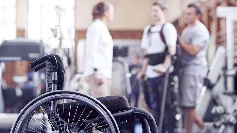
Biologists and engineers are working together to dismantle the assumption that spinal cord injury is permanent. Image (licensed) from Adobe Stock
Electrical engineers are playing a significant role in these efforts, perhaps viewing the problem as a new signal-integrity challenge requiring innovative solutions. Their tools aren't drugs or rehabilitation protocols alone: they're electrode arrays, skin-surface sensor grids, tiny robots with magnetoelectric nanoparticles, and brain implants. And they're all at different stages of proving that the old assumption—"once the cord is broken, it stays broken"—is no longer technically true.
Electrode Arrays Restore Spinal Cord Communication
The spinal cord is essentially a two-lane communication link: one lane carries motor commands down from the brain; the other carries sensory feedback back up. A complete lesion physically severs both lanes. You can't move your legs, and you can't feel where they are.
A team at Brown University used electrode arrays above and below the injury site to bridge the severed connection in both directions. In three patients with complete lower-limb paralysis, the electrodes enabled stimulation of the spinal cord below the lesion, allowing patients to partially regain muscle control. Above the lesion, the same arrays allow patients to sense their leg position. That bidirectional restoration had never been demonstrated before in complete spinal cord injury.
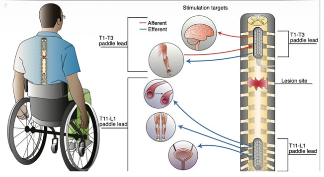
Electrode arrays above and below the injury simultaneously stimulate both sensory and motor pathways. Image used courtesy of Brown University
To find the right stimulation parameters, the team didn't rely on weeks of manual trial and error. Instead, they framed the problem as a patient-specific parameter optimization. Each patient's neural response to stimulation served as the feedback signal, and a deep learning model trained on that individual's data automatically converged on optimal settings.
Participants used a "DJ board," a physical controller with knobs and sliders, to manually explore stimulation patterns while aiming to achieve specific leg movements. This human-in-the-loop exploration generated the labeled dataset the model required. A deep learning algorithm then analyzed this data to automatically identify optimal stimulation settings in under 20 minutes, a task that would otherwise require weeks of manual trial and error.
Tracking Coordination Between Neural Signals
While stimulation can activate muscles, it doesn't necessarily restore a patient's ability to move their limbs. For that, a precisely timed sequence of neural signals must be exchanged between brain and muscles to produce smooth, purposeful movement.
To better understand the coordination of neural signals, researchers at KTH Royal Institute of Technology in Sweden placed a high-density 2D grid of electrodes on the skin. The grid isolates and tracks individual motor unit action potentials—the smallest functional signals in muscle. Unlike conventional methods, which reflect the superposition of many units and obscure timing information, this approach isolates each unit's contribution separately.
In people with incomplete spinal cord injuries, the team found that even when patients tried very lightly (at just 20% of their maximum strength), motor units fired out of sync. The result was shaky, unstable force output, producing a direct quantitative measure of the balance difficulties these patients experience. However, when the patient exerted greater effort, the nervous system overcompensated by sending louder, less refined signals.

One of the study participants exerts force with their calf muscles while sensors measure electrical activity. Image used courtesy of KTH Royal Institute of Technology
While this method is not a treatment, it offers insight into the function of residual neural pathways. Such findings may one day provide a non-invasive way to measure recovery in response to other spinal cord injury therapies. It requires no surgery and no implants. Instead, a simple skin-surface grid provides clinicians with an assessment tool to determine whether neural coordination, not just muscle strength, is actually improving.
Microrobots That Build New Nerves From the Outside
To address cases in which nerves are simply gone, researchers at ETH Zurich and the University of Zurich have developed tiny robots, guided by magnets from outside the body, that can rebuild them. Their construct, the NPCbot, is about six micrometers in diameter—roughly one-tenth the width of a human hair.
Each bot is a special type of living neural cell bound to a magnetoelectric core-shell nanoparticle (cobalt ferrite inside, barium titanate outside). The bots are injected directly into the injury site, where an external magnetic field then guides them precisely to the target tissue.
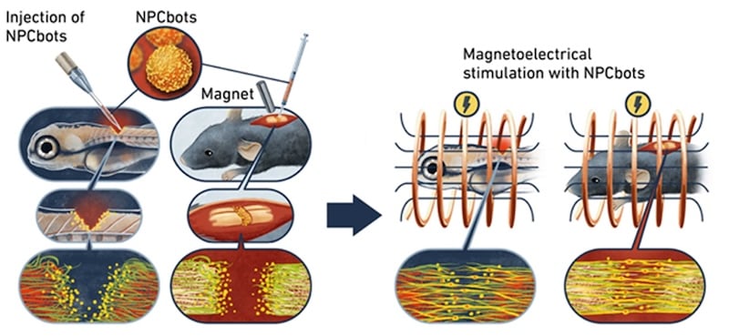
Microrobots injected near the injury are guided and activated by an external magnet to grow new nerve cells. Image used courtesy of ETH Zurich
Here's the engineering trick: apply a low-frequency alternating magnetic field from outside the body in the kilohertz range, at amplitudes on the order of tens of millitesla. The inner core deforms (magnetostriction) in response. That mechanical strain couples across the core-shell interface and transfers to the outer shell, which converts it into an electrical voltage through the piezoelectric effect.
The result is a localized electrical stimulus delivered right at the cell surface, triggering the stem cell to differentiate into a neuron. The entire stimulus chain is contactless: no leads, no implanted electrodes, no genetic modification.
Conventional stem cell transplants face a well-documented limitation: prior studies report that only about 5% of transplanted cells survive long enough to be therapeutically useful, and that differentiation at the target site is unreliable. The NPCbots, by contrast, are magnetically guided to the injury site and wirelessly activated through the same field used for navigation—effectively combining magnetic steering and on-site electrical stimulation into a single external control signal.
In zebrafish, the animals recovered nearly normal swimming behavior within three days. In mice, nerves reconnected across the injury zone after 28 days, and the animals moved measurably better.
The World's First Commercially Approved Brain-Computer Interface
Shanghai-based Neuracle Technology, together with researchers at Tsinghua University, has developed NEO—a coin-sized brain implant that rests on the dura mater, the tough membrane covering the brain. When a patient imagines grasping an object, NEO's electrodes detect epidural electrocorticography (ECoG) signals from the motor cortex, capturing the neural signature of motor intention. Then, a decoding algorithm identifies the intent in real time and wirelessly commands a pneumatic glove on the patient's hand to execute the movement.
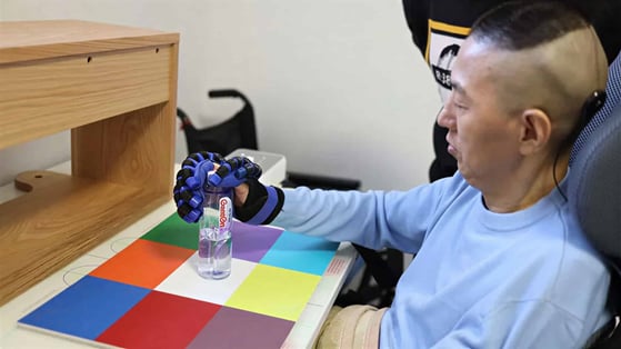
A paralyzed patient uses the NEO brain implant to grasp a bottle. Image used courtesy of Tsinghua University and Neuracle Technology
The decoder, built on Riemannian geometry, first extracts spatio-spectral features from the signal's covariance structure. A Hidden Markov Model then tracks the temporal dynamics of continuous grasping intent and translates it in real time into a wireless command. That command drives the pneumatic glove on the patient's hand. Using this approach, one patient with a complete C4 cervical injury—paralyzed in all four limbs for over a decade—recovered the ability to grasp objects and drink independently.
The implant has no battery. Instead, power and bidirectional data are transferred wirelessly through inductive coupling, in which an external coil worn against the scalp communicates with an internal coil through the skin. The device functions only when this external coil is being worn.
Since October 2023, Neuracle has run 36 clinical trials. In March 2026, China's National Medical Products Administration granted NEO commercial approval, making it the first invasive brain-computer interface approved for use outside clinical trials. The approval currently covers patients aged 18 to 60 with cervical injuries who retain some upper-arm function, though it is limited to China.
Engineering Challenges Ahead
Designing circuits and devices for spinal cord injury comes with unique challenges. Electrode degradation and tissue encapsulation over years of implantation can compromise the long-term reliability of these solutions. Efficient inductive coupling through tissue and heat dissipation can also undermine wireless power and data transfer. Scaling presents another challenge: moving from three patients to three hundred—each with different injury profiles, ages, and real-world challenges—introduces significant hurdles.
The honest caveat is that most of these studies are still in animal models or small patient cohorts. But with the pace of this research accelerating, biologists and engineers are working together to dismantle the assumption that spinal cord injury is permanent.